威斯尼斯人(wns615app-百度VIP认证)-Wikipedia

Product Center
描述
描述
ABOUT
US
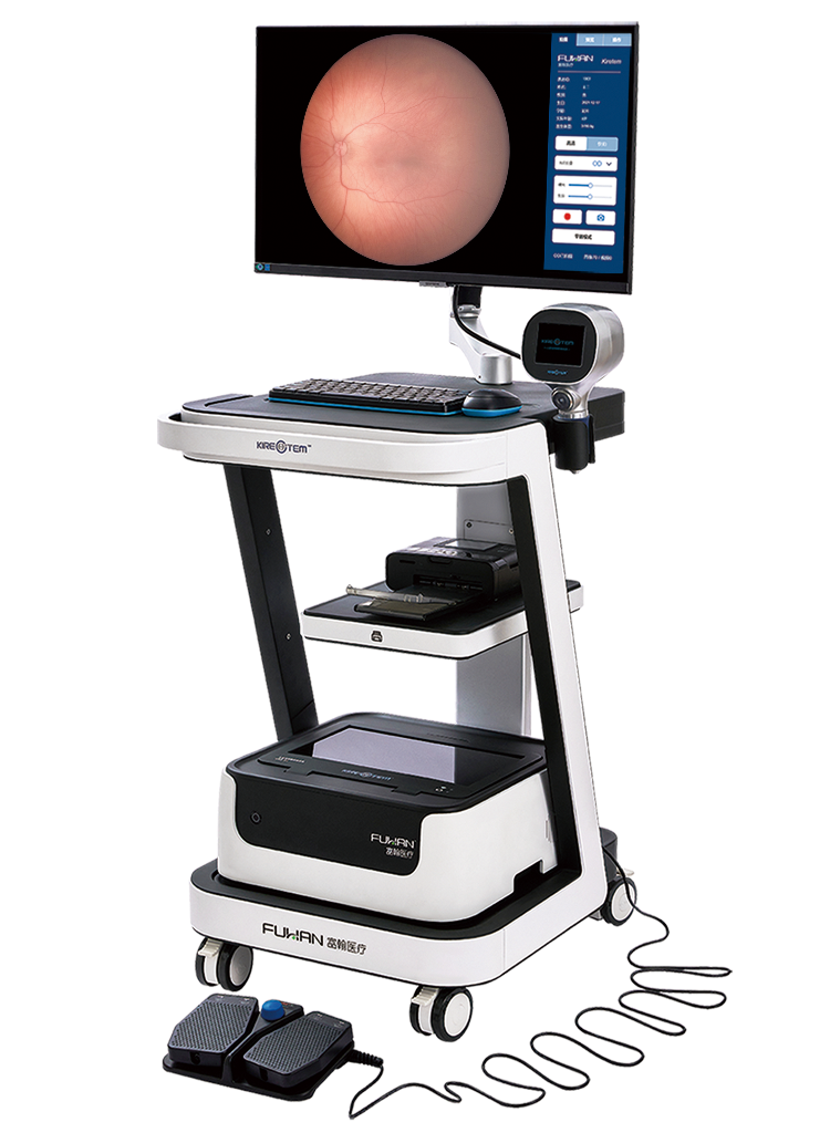
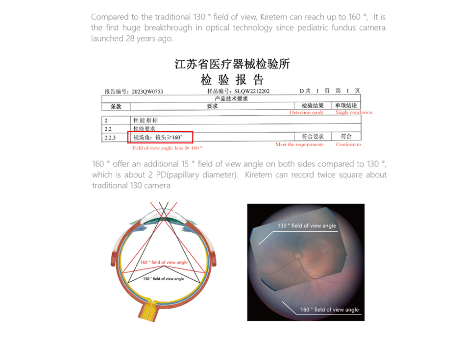
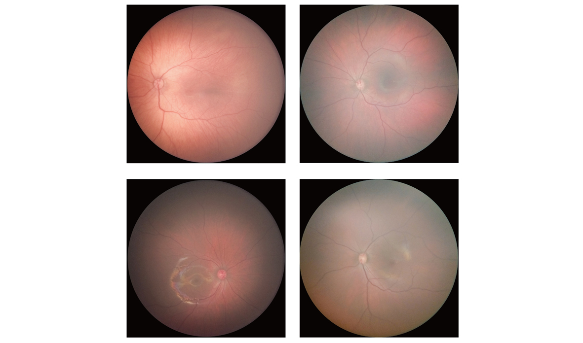
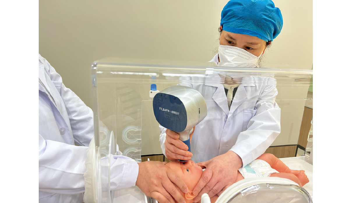
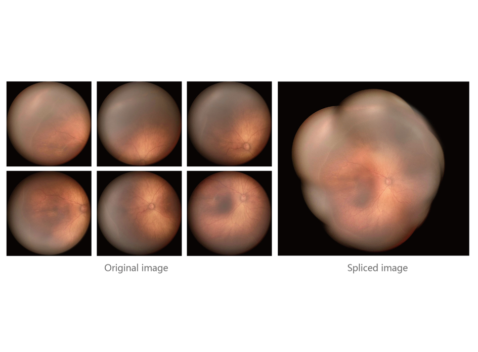
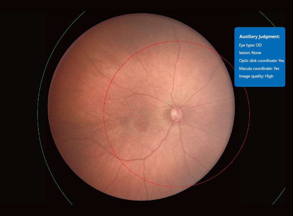
Kiretem Ophthalmic Wide-Angle lmaging System is a very popular equipment for screening fundus disease of infants and children in clinical practice, and it is an objective and non-injury-free examination method. The operation is simple and fast, the field of view is large, and the image quality is high, which is conducive to early detection of a variety of neonatal congenital eye diseases and fundus lesions, and provides a reliable basis for early treatment and early intervention of neonatal eye diseases.
Kiretem Ophthalmic Wide-Angle lmaging System KRS-01
描述
01
Exclusive 160 °field of view
   Effective prevention of missed diagnosis: Kiretem 160° provides an additional 15° field of view on each side compared to the traditional 130°, about 2 PD (optic disc diameter), fundus imaging range is wider, and peripheral retinal images are easier to obtain. When shooting the same part, the imaging range of the 160° lens is about twice that of the 130° lens, which can effectively reduce the missed diagnosis of peripheral retinal diseases.

  Easier to operate:The Kiretem handle has a smaller deflection and is easier to operate during periretinal image acquisition, reducing the learning curve for healthcare providers.
描述
02
True color high-definition imaging 
   Restore the true color of the retina: Kiretem use a 13-million-pixel HD CMOS camera, support static shooting and image recording functions, present higher quality and clearer retinal local details, restore the true color of the retina, reduce the misdiagnosis caused by blurred retinal images.
描述
03
7mm Minimum lens diameter
  7mm small-diameter lens is more suitable for the examination of premature infants with small palpebral cleft, reducing the medical risk caused by the lens being too large to perform scheduled screenings on children.

  The aperture of the 7mm small-diameter lens is only 5.2mm, and the aperture diameter of similar products is more than 6mm. Kiretem small aperture lens is more suitable for small pupil imaging, for clear imaging, a dilated pupil diameter ≥4.5mm is required.
描述
04
Single handle wireless mode 
   The 500g handle, extremely portable, single handle to complete registration, inspection and printing.
描述
05
One click panoramic viewing 
   Automatically capture multiple high-quality fundus images of different directions, splice them based on blood vessel morphology and anatomical positioning of macula and optic disc, and present the whole fundus image. It makes the film reading diagnosis more intuitive and accurate, and provides help for ROP partitioning.
描述
06
AI assisted judgment system
   Help doctors accurately locate the location of optic disc and macula, judge whether the photo is clear, whether there is an abnormality in the fundus, further distinguish the staging of the disease, and solve the problem that primary medical units can not carry out pediatric eye screening work due to the lack of fundus disease physicians.